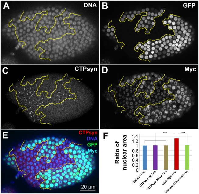
Fig 5. CTPsyn knockdown supresses Myc-induced overgrowth phenotype in Drosophila follicle cells.
(A-E) The nuclei of cells both overexpressing Myc and knocking down CTPsyn (UAS-Myc, CTPsynRNAi) are marked by GFP (B, outlined by yellow lines in A-E). Myc overexpression is verified by immunostaining with an antibody against Myc (D). CTPsyn knockdown is verified by immunostaining with an antibody against CTPsyn (C). Note that no cytoophidia are detectable in the clonal cells even when Myc is overexpressed. (F) Quantification of mid-stage follicle cells shows that Myc overexpression (UAS-Myc) alone increases nuclear size significantly. Follicle cells in UAS-Myc, CTPsynRNAi cells have similar nuclear size compared to non-clonal (nc) cells. CTPsynRNAi or CTPsyn overexpression show no significant difference in nuclear area (see S10 and S11 Figs for representative images). Quantification represents the mean nuclear areas from > 50 cells in > 3 egg chambers per genotype. ***P<0.001. n.s. = not significant. Error bars show SEM.

