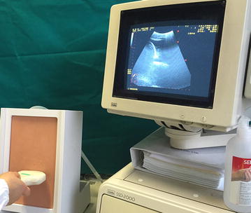
Fig. 1.

The thoracentesis simulator ‘Ultrasound Thoracentesis Model THM-30’ developed by SIMULAB, Seattle, USA. It features a partial torso with anatomical landmarks, such as skin texture, ribs, and a fluid reservoir. Its simulated lung is seen as an echogenic structure with an inflating mechanism to adjust the size of the pleural effusion
