
Figure 3.
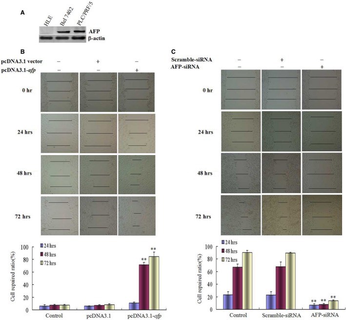
Effects of AFP on wound heal of human liver cancer cells HLE and Bel 7402 cells. (A) Expression of AFP in HLE and Bel 7402 cells was detected by Western blotting, PLC/PRF/5 cells were used as a positive control of AFP expressed. (B) HLE cells were transfected with pcDNA3.1‐afp vectors for 24, 48, 72 hrs, respectively; wound healing of the cells were observed by microscopy; low columnar graph showed repaired‐ratio of the cells; **P < 0.01 versus control and pcDNA3.1‐vectors groups. (C) Bel 7402 cells were transfected with AFP‐siRNA vectors for 24, 48, 72 hrs respectively; wound healing of the cells were observed by microscopy; low columnar graph showed repaired‐ratio of the cells; **P < 0.01 versus control and scramble‐siRNA groups. We performed three independent experiments.