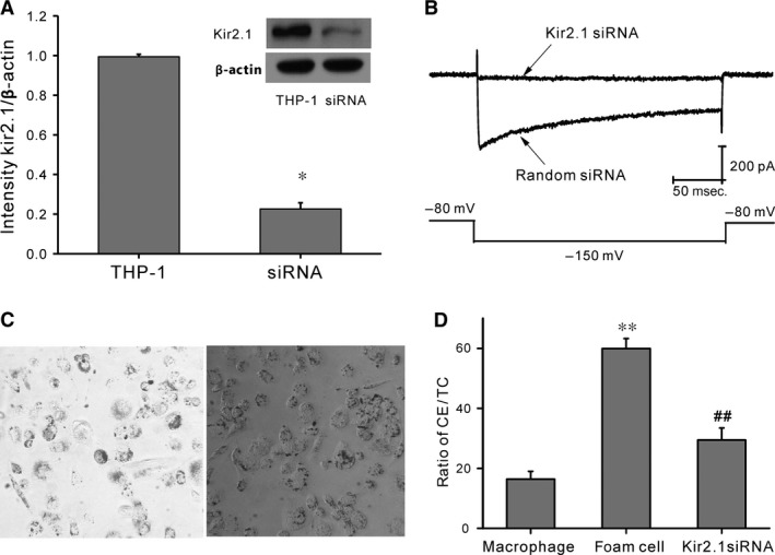
Figure 4.

Kir2.1 silencing using Kir2.1 siRNA. (A) Typical protein expression gels (top) and histogram (bottom) showing Kir2.1 protein expression following transfection of Kir2.1 and scrambled siRNAs. *P < 0.05, scrambled siRNA versus Kir2.1 siRNA. (B) Representative traces of Kir2.1 currents from THP‐1 cells treated with Kir2.1 siRNA (top) or scrambled siRNA (bottom). (C) Oil red O staining of THP‐1–derived macrophages treated with 30 mg/l ox‐LDL (×100). Left: Control group; right: Kir2.1 siRNA group. (D) Histogram showing the ratio of intracellular CE in macrophages, foam cells, and Kir2.1 siRNA‐transfected macrophages. **P < 0.01 versus macrophages; ##P < 0.01 versus foam cells. Values are the mean ± SE.
