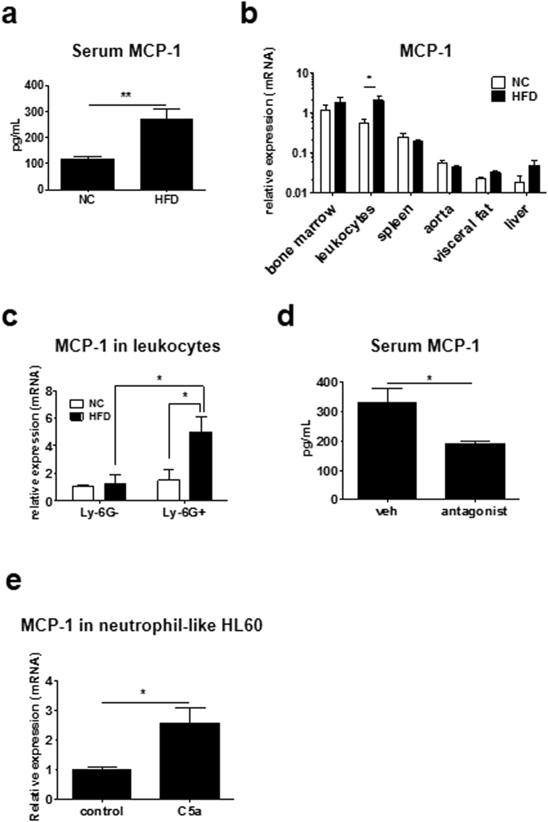
Figure 4. HFD upregulated MCP-1 expression in neutrophils.
(a) HFD for 4 weeks significantly increased MCP-1 levels in sera of mice (n = 8 and 8, respectively). Data are presented as the mean ± SEM. **P < 0.01 by unpaired; 2-tailed Student’s t test. (b) MCP-1 mRNA levels in various tissues including bone marrow (n = 8 and 8, respectively), leukocytes (n = 4 and 5, respectively), spleen (n = 7 and 8, respectively), aorta (n = 6 and 8, respectively), visceral fat (n = 8 and 8, respectively) and liver (n = 8 and 7, respectively), of mice fed HFD or NC, involving 4 independent experiments. HFD significantly increased the MCP-1 level in circulating leukocytes. Data are presented as the mean ± SEM. *P < 0.05 by two-way ANOVA with Bonferroni’s post test. (c) HFD increased MCP-1 mRNA levels in neutrophils such as Ly-6G+ leukocytes, but not Ly-6G− leukocytes (n = 3/each group), involving 4 independent experiments. Data are presented as the mean ± SEM. *P < 0.05 by two-way ANOVA with Bonferroni’s post test. (d) MCP-1 level in sera was measured by ELISA when C5aR antagonist was administrated in wt mice fed HFD for weeks. C5aR antagonist significantly decreased serum MCP-1 levels increased by HFD (n = 4 and 4, respectively). Data are presented as the mean ± SEM. *P < 0.05 by unpaired; 2-tailed Student’s t test. (e) mRNA level of MCP-1 in neutrophil-like HL-60 treated with 3nM recombinant C5a. C5a significantly increased MCP-1 expression level compared to control (n = 5 and 5, respectively), involving 4 independent experiments. Data are presented as the mean ± SD. *P < 0.05 by unpaired; 2-tailed Student’s t test.

