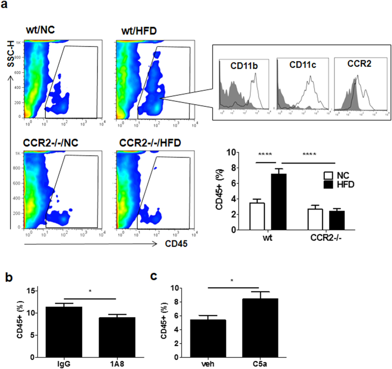
Figure 5. Neutrophils contributed to intimal CD45-positive leukocytes in vessels.
(a) Flow cytometry for leukocytes in femoral artery of wt mice or CCR2−/− mice fed NC or HFD. CD45+ cells were gated by the square (upper). Leukocytes in the intima significantly increased in wt mice fed HFD compared with NC. However, the CD45-positive cells did not change in CCR2−/− mice between HFD and NC (n = 7/each group). These leukocytes were positive for CD11b, CD11c, and CCR2, involving 8 independent experiments. An isotype control antibody was used as the negative control (gray filled). Data are presented as the mean ± SEM. ****P < 0.0001 by two-way ANOVA with Bonferroni’s post test. (b) Flow cytometric analysis for single-cell suspension of femoral arteries of wt mice fed HFD for 4 weeks when neutrophils in peripheral blood were depleted by specific antibody. Neutrophil depletion significantly decreased the intimal leukocytes in the femoral artery (n = 6 and 6, respectively). Data are presented as the mean ± SEM. *P < 0.05 by unpaired; 2-tailed Student’s t test. (c) Flow cytometric analysis for single-cell suspension of the femoral arteries of wt mice treated with C5a for 4 weeks. C5a significantly increased leukocytes in the femoral artery (n = 8 and 7, respectively). Data are presented as the mean ± SEM. *P < 0.05 by unpaired; 2-tailed Student’s t test.

