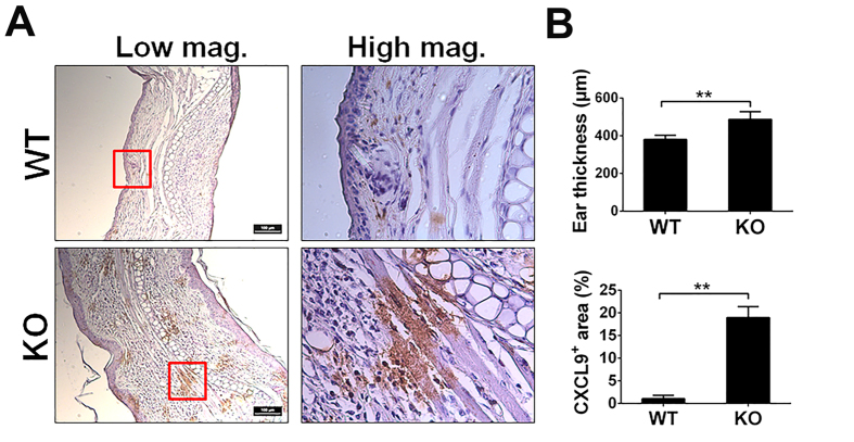
Figure 4. The expression of CXCL9 in ear of OXA-induced contact dermatitis.
Contact dermatitis was induced on the ear of WT and PGRN KO mice by OXA (n = 6 group). After two weeks, mice were sacrificed and ear tissues were fixed for histology analysis. Ear thickness and CXCL9 expression were examined. (A) Immunohistochemistry staining of CXCL9 in ear. Tissue from WT and PGRN KO mice were stained with CXCL9 antibody and expression of CXCL9 is shown in brown. (B) Quantification of ear thickness (upper panel) and CXCL9 (lower panel) expression. Ear thickness is measured at five different positions on each ear sample and the average ear thickness for each mouse was calculated by Image J based on the scale bar. The data is presented as averaged thickness leveled across 6 mice per group. For of CXCL9 staining quantification, five images were taken for each sample under 40X objective lens field. And the positive staining were circled and calculated by Image J. The data is represented as ratio of CXCL9 positive area to total area of 40X objective lens field. One-way ANOVA was used for statistical analysis (**p < 0.01).

