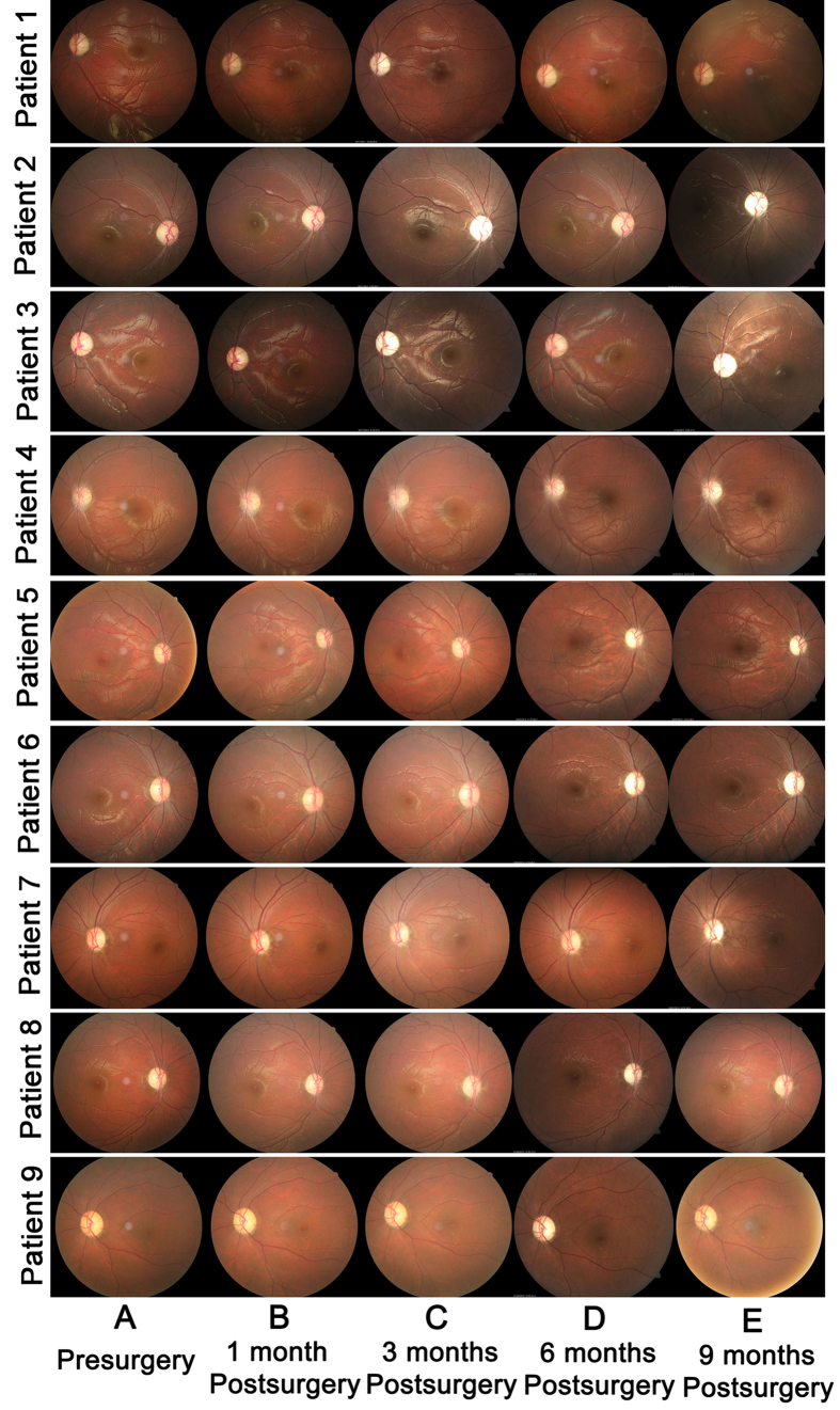
Figure 1. Retinal appearance and morphologic features before and after intravitreal injection of rAAV2- ND4 in the nine patients.
(A) Representative 30° fundus photographs taken with a NIDEK Auto Fundus Camera show the disk and macula of injected eyes of patients before intravitreal injection. The retinal structure of the nine patients was normal. (B to E) 30° fundus photographs show the disk and macula of injected eyes of the nine patients at months 1, 3, 6, and 9 after intravitreal injection, respectively. No apparent retinal abnormality was found.

