
Figure 2.
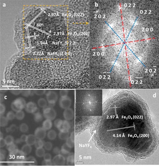
HRTEM studies of NPs: (a) HRTEM images of Fe3O4@NaYF4(Yb, Tm); (b) fast Fourier transform of the selected area in part a, showing two sets of diffraction patterns. The diffraction pattern marked in blue belonged to cubic Fe3O4, and the one marked in red was assigned as cubic NaYF4; (c) high angle annular dark field image of Fe3O4@NaYF4(Yb, Tm), showing the Z contrast difference between the shell and core of particles induced by a slightly higher average atomic number in the shell after doping with heavy atoms Yb and Tm; (d) HRTEM image revealed the core–shell structure of NP Co0.16Fe2.84O4@NaYF4(Yb, Er). Atomic lattice fringes 2.97 and 4.14 Å corresponded to (022) and (200) planes of Fe3O4, respectively. The inset is a fast Fourier transform of the micrograph.