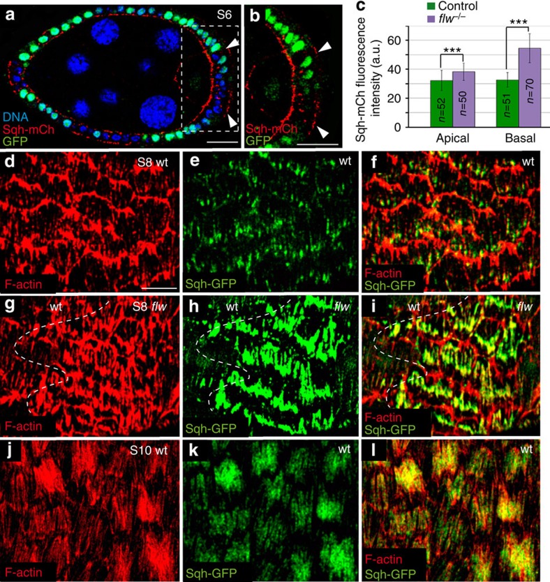
Figure 2. Loss of flw enhances basal recruitment of actin and myosin.
(a) Sagittal plane of a mosaic S8 follicle expressing Sqh-mCherry (red) and containing flw mutant clones stained for anti-GFP (green) and the nuclear marker TO-PRO-3 (blue). flw mutant FCs (labelled by the absence of GFP, arrowheads) show higher levels of basal Sqh-mCh than controls (GFP+). (b) Magnification of the white box in a. (c) Quantification of the apical and basal levels of Sqh-mCh in control and flw mutant FCs. The statistical significance of differences was assessed with a t-test, *** P value<0.0001. All errors bars indicate s.d. (d,l) Surface view of control S8 (d–f), mosaic S8 (g–i) and control S10 (j–l) egg chambers expressing Sqh-mCherry, stained for anti-GFP (green) and Rhodamine-Phalloidin to detect F-actin (red). (g–i) The actomyosin fibers of flw mutant FCs exhibit higher levels of F-actin and myosin compared with controls (d–f). Scale bar, 10 μm. Mean of n>42 FCs, assessed over five independent experiments.

