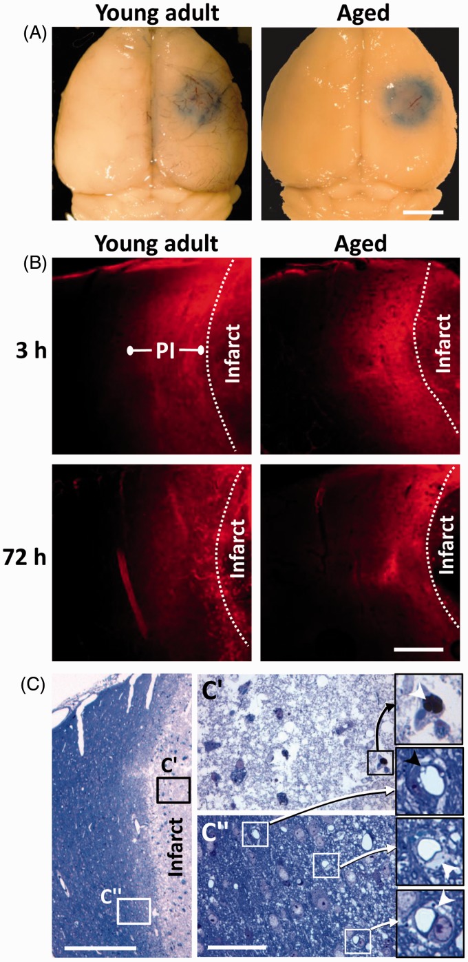
Figure 1.
Focal ischemic stroke leads to a long-lasting increase in vascular permeability in the peri-infarct cortex of young adult and aged mice. (a) Whole-brain brightfield images showing Evans blue dye extravasation around the cerebral infarct 3 h after initiating photothrombosis. Scale bar = 2 mm. (b) Representative confocal images (Z projection of 10 planar images taken in 4 µm steps) showing the halo of Evans blue fluorescence around the cerebral infarct. This halo of diffuse fluorescence usually extends ∼300–500 µm from the infarct border, which we refer to as “peri-infarct” (PI) cortex. Note that cortical areas more distant to the infarct exhibit little to no fluorescence. Scale bar = 200 µm. (c) A toluidine blue stained 0.5 µm thick section showing the infarct core (c′) and peri-infarct region (c″) 72 h after stroke in an aged mouse. The vessels inside the infarct core were either plugged (see arrowhead within inset from c′) or unrecognizable. Open perfused microvessels were evident in the peri-infarct zone (c″). Close inspection reveals vacuolated endothelium (black arrowhead) and swollen astrocyte endfeet (see white arrowheads within insets from c″). Scale bar = 400 µm for c and 50 µm for c″.

