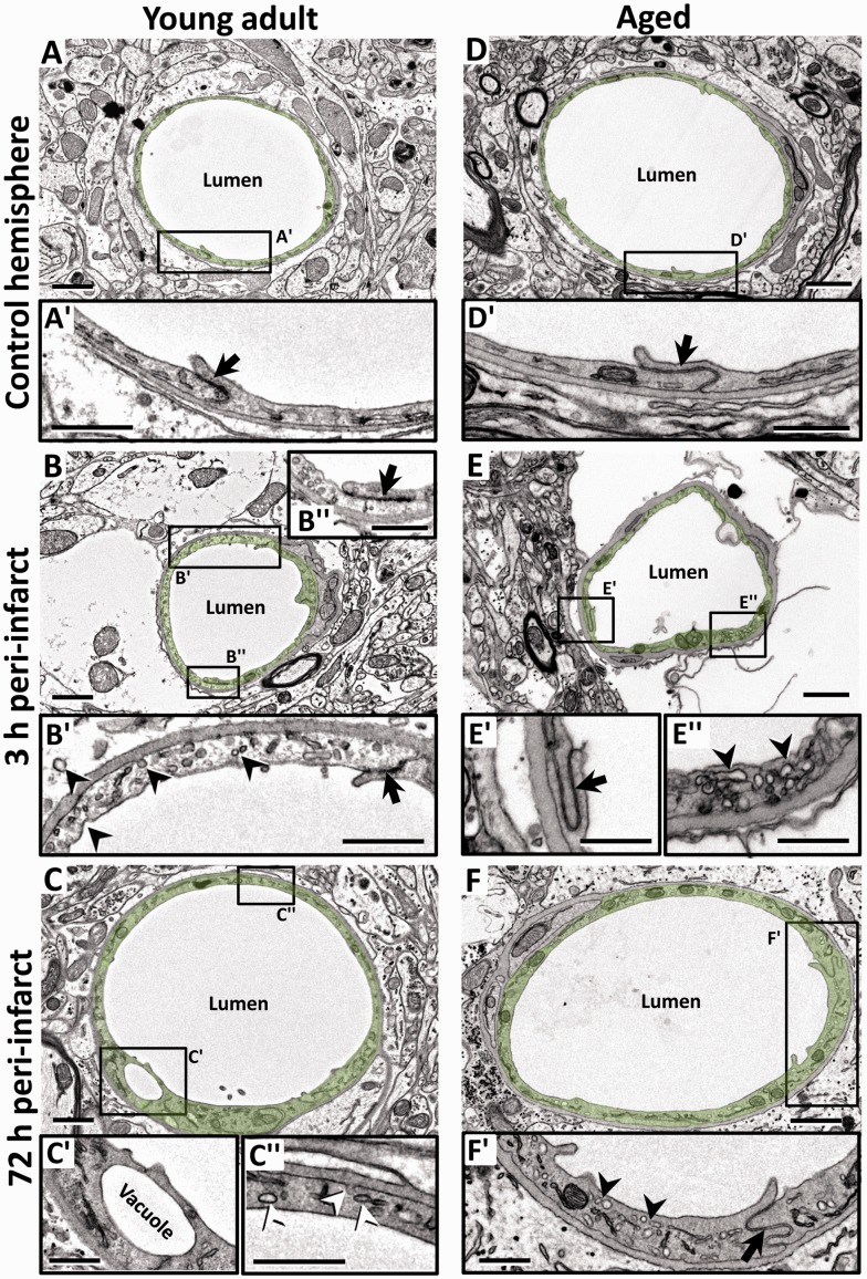
Figure 2.
Focal ischemia induces endothelial swelling and upregulates caveolae and vacuole formation in peri-infarct capillaries in young adult and aged mice. (a, d) Electron micrographs (EMs) of capillaries in the nonischemic contralateral hemisphere in young adult (a) and aged (d) mice. Endothelium is lightly shaded green. Insets (a′ and d′) below each image show a relatively thin endothelial layer and intact TJs (arrow) at both ages. (b, e) Representative capillaries in peri-infarct cortex of young adult (b) and aged (e) mice 3 h after stroke. Insets (b′ and e″) show the slightly swollen endothelium that was densely packed with putative caveolae-like vesicles (arrowheads) on the luminal and abluminal sides. However, TJs (black arrows in b″ and e′) was intact at both time points. (c, f) EMs from young adult (c) and aged (f) mice showing dilated peri-infarct microvessels 72 h after stroke. At this time point, the endothelium appeared more swollen and was still laden with caveolae-like vesicles (c″ and f′) and the occasional vacuole (c′). TJs were generally intact (f′). Scale bar = 1 µm for a to f. Scale bar = 0.5 µm for insets a′ to f′.

