Figure 4.
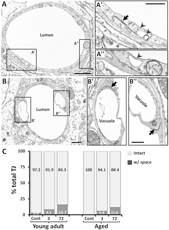
Vacuole and vesicle formation in relation to the tight junction. (a) EM of a peri-infarct capillary 3 h after stroke in a young adult mouse. On occasion, a membrane-bound space was evident at the TJ (black arrow in a′), perhaps due to the pooling and fusion of transcytotic vesicles (arrowheads in a′). However, these vacuoles could be found in parts of the endothelium not associated with TJ (a″). (b) Of note, vacuoles often formed in endothelium adjacent to the TJ (b′–b″). (c) Histogram shows the percentage of TJs that were completely intact or had a space present in young adult and aged mice. Although there were slightly more TJs with spaces after stroke (e.g. at 72 h), they were relatively infrequent. Percentages were based on the analysis of 34–43 TJs per group. Three mice were analyzed in each group. Scale bar = 1 µm for a and b and 0.5 µm for a′–b″.

