Figure 5.
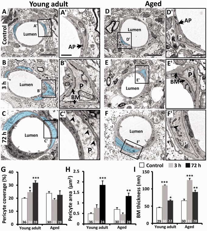
Effects of ischemia on pericyte area, basement membrane thickness, and pericyte coverage of the vascular endothelium. (a to f) EMs of microvessels in young adult (a to c) and aged mice (d to f) in the contralateral control hemisphere or peri-infarct cortex 3 and 72 h after stroke. Pericytes are highlighted in blue. (a′ and d′) In control microvessels, pericyte processes (P) were encapsulated with a thin basement membrane (BM) and showed typical features such as an adhesion plaque (AP) at endothelial cell contact points. Three hours after stroke (b, e), the basement membrane thickened considerably and pericyte processes appeared slightly swollen (b′, e′). At 72 h, pericyte swelling increased along with its coverage of the endothelium in young adult mice (c′). However in aged mice, pericytes appeared swollen (f′) but do not show any change in their coverage of the endothelium (g). Quantitative analysis indicated that pericyte coverage of the endothelium (g) and cross-sectional area (h) increased significantly in peri-infarct cortex at 72 h in young adult mice. (i) Thickness of the basement membrane region increased significantly in both age groups at 3 h recovery and remained elevated in aged mice. The number in each histogram bar represents the number of capillaries analyzed per group, sampled from three to five mice per group. Scale bar = 2 µm for a to f. Scale bar for insets a′–f′ = 0.5 µm. *p ≤ 0.05, ***p ≤ 0.001 relative to controls.

