Figure 6.
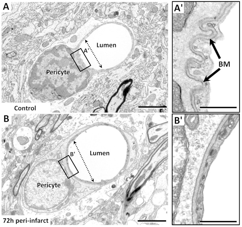
Examples of a pericyte wrapping around a microvessel. (a and b) EMs of a pericyte in the control hemisphere (a) or in peri-infarct cortex 72 h after stroke (b). Note that in the control microvessel, the endothelium and basement membrane were corrugated next to the pericyte soma (a′), and the lumen proximal to the soma was narrower than that distally, suggestive of a contractile function. Seventy-two hours after stroke when microvessels tended to be dilated, the corrugated appearance of the basement membrane was absent (b′), and the lumen of the vessel was larger. Scale bar = 2 µm for (a)–(b). Scale bar for insets a′–b′ = 0.5 µm.

