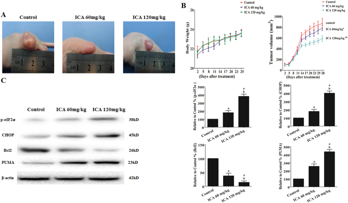
Figure 9. Effect of ICA on EC109 tumor xenografts in vivo.
(A) Photographs showing tumor xenograft morphologies in each group. (B) Change in body weight of the mice. A tumor growth curve was drawn from the tumor volumes and treatment duration. (C) Representative Western blot results of p-eIF2α, CHOP, Bcl2, and PUMA are shown. Membranes were re-probed for β-actin expression to show that similar amounts of protein were loaded in each lane. The results are expressed as the mean ± SD; n = 6. aP < 0.05 vs. the control group; bP < 0.05 vs. the 60 mg/kg ICA-treated group.

