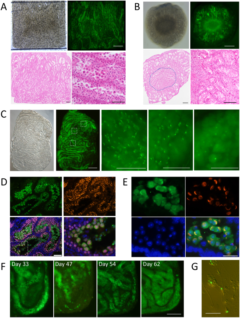
Figure 2. Progression of spermatogenesis in the microfluidic device.
(A) Testis tissue was spread flat in the tissue chamber. GFP expression was observed in most tubules, observed on culture day 42. Histological examination confirmed that most seminiferous tubules contained germ cells in the meiotic phase (HE). (B) Tissues cultured with the AG method showed GFP expression in a donut-like fashion, with the central region degenerated. HE staining histology demonstrated such changes. The circled area is magnified in the right lower panel showing the degenerative tubules. (C) Testis tissue of 4.5 dpp mice cultured for 38 days in the device. Central areas of the 3 dashed rectangles are enlarged in the right panels, showing that each portion is in a different stage of spermatogenesis. (D) Immunohistochemical examination with antibodies to GFP (green) and SCP3 (red), counterstained with Hoechst (blue). The bottom left panel is the merged image. The dashed rectangular area is enlarged in the bottom right panel. The GFP(−)SCP3(+) cells were observed surrounding the GFP(+)SCP3(+) cells inside, along with inner-most GFP(+)SCP3(−) cells, confirming that Acr-Gfp expression starts in the mid-pachytene stage of meiosis and protein remains in round spermatids. (E) Lectin PNA staining (red) demonstrated that GFP-dots and -caps corresponded to the acrosome structure. (F) A single spot, time-course observation revealed that GPF-positive cells repeated emergence and disappearance. (G) Sperm with GFP-acrosomes and flagella were found in the suspension produced by tissue dissociation. Scale bars: 500 μm (A,B upper, C left), 200 μm (A,B lower, F), 100 μm (D), 50 μm (C right), 20 μm (E,G).

