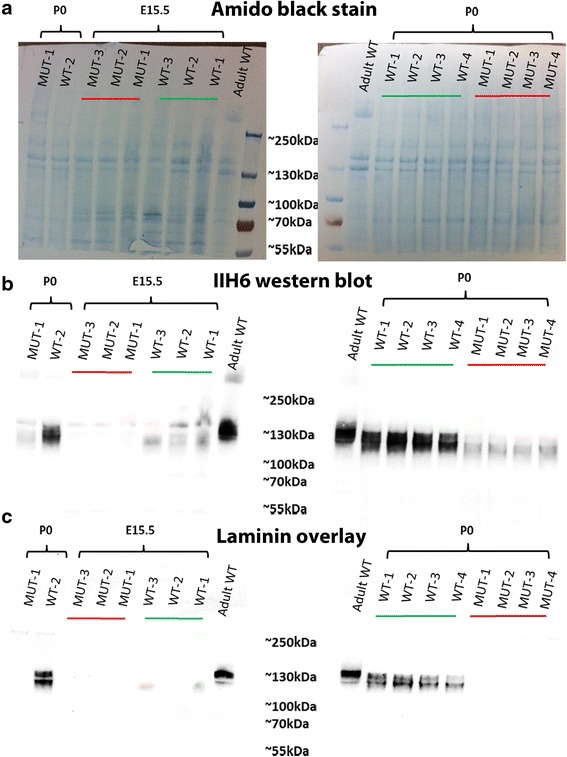
Fig. 3.

IIH6 Western blot and laminin overlay at E15.5 and P0. a Shows an amido black stain of the transfer membrane indicating equivalent loading in all lanes. b Western blot for IIH6 in the hindlimb muscle at E15.5 and P0, the relative molecular mass of α-dystroglycan in the wild type is reduced compared to the adult mouse muscle and only residual IIH6 labelling was observed in FKRPKD. c At both E15.5 and P0, α-dystroglycan was able to bind laminin in the wild type and no binding was observed in the FKRPKD
