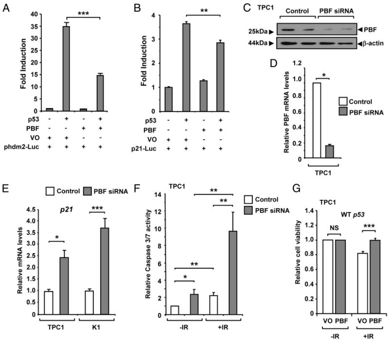
Figure 4.
PBF inhibits p53 transcriptional activity. H1299 cells were transfected with p53 luciferase reporter plasmids for either (A) hdm2 (phdm2-Luc) or (B) p21 (p21-Luc), as well as p53 and PBF expression vectors, or vector only (VO) as indicated. Luciferase activity was measured 24 hours post-transfection. C-D, TPC1 cells were transfected with either PBF-specific or control siRNA at a final concentration of 100 nM. PBF expression was assessed by Western blotting and qRT-PCR analysis as shown. E, Relative mRNA levels of p21 mRNA in TPC1 and K1 cells transfected with either PBF-specific or control siRNA for 48 hours at a concentration of 100 nM. F, Analysis of caspase-3/7 activity in TPC1 cells transfected with either PBF-specific or control siRNA for 48 hours and then irradiated with a 15 Gy dose (+IR) or untreated (−IR). Normalized mean caspase-3/7 values ± SE are shown from 4 independent experiments. For each experiment caspase-3/7 activity was determined from n = 5 per condition at 24 hours postirradiation. G, TPC1 cells were transfected with either VO or PBF for 24 hours and either untreated (−IR) or irradiated (+IR) with a 15 Gy dose. Cells were then replated and viability measured after 24 hours. Data presented as mean ± SE from four independent experiments. *, P < .05; **, P < .01; ***, P < .001; NS = not significant.

