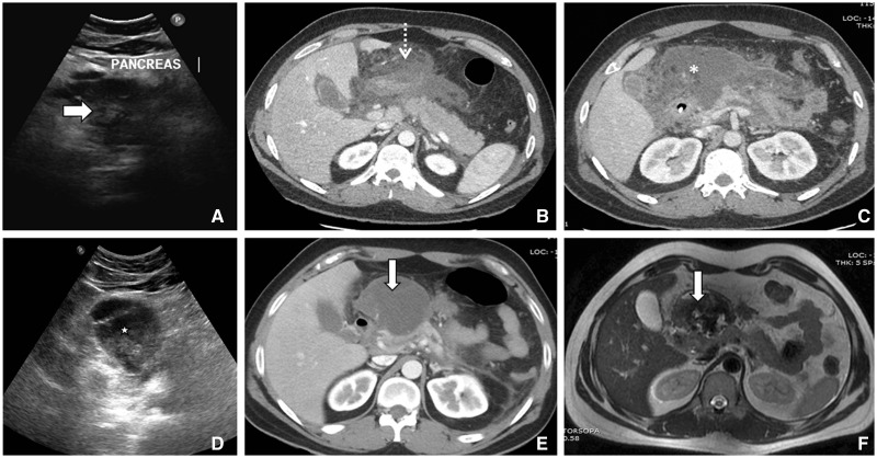
Figure 5.
Serial images of a 26-year-old male presenting with acute necrotizing pancreatitis. (A) First ultrasound showing bulky heteroechoic pancreatic (arrow) echotexture. (B) Axial contrast-enhanced CT, showing acute necrotic collection (dashed arrow) with absence of a well-defined wall involving predominantly the peripancreatic region and neck of pancreas. (C) Subsequent axial CT scan around 15 days later, showing increasing heterogeneity (asterix) with areas of ill-defined wall. (D) Ultrasound 4 weeks later than the first ultrasound, showing walled-off necrotic collection (star) in the form of well-defined wall with coarse internal echoes. (E) Axial CT and (F) T2-weighted MR image showing walled-off necrosis (arrow).

