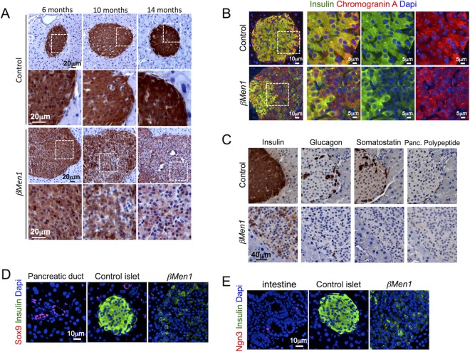
FIG 2.
Progressive loss of insulin expression in βMen1 tumor β cells. (A) Immunohistochemical analysis of insulin expression at the indicated ages. (B) Analysis of chromograninA and insulin coexpression. Representative pictures of double-immunofluorescent staining from 14-month-old βMen1 and control mice are shown. (C) Immunohistochemical staining of insulin, glucagon, pancreatic (Panc.) polypeptide, and somatostatin in 14-month-old pancreas serial sections. (D and E) Analysis of Sox9 and Ngn3 expression in 14-month-old Ins− βMen1 tumor lesions. Note that pancreatic ducts and intestinal tissues present on the same pancreatic sections were used as internal controls expressing Sox9 and Ngn3, respectively.

