FIG 1.
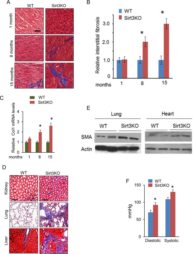
Reduced SIRT3 levels contribute to tissue fibrosis. (A) Comparative study of fibrosis in hearts, stained with Masson's trichrome stain (blue), of Sirt3-KO mice of different age groups and their age-matched wild-type (WT) controls. (B and C) Quantification of cardiac fibrosis and Col1 mRNA levels in Sirt3-KO and WT mice of different age groups. Values are means ± standard errors (n = 4). *, P < 0.01. (D) Tissue sections of 15-month-old WT and Sirt3-KO mice, stained to detect fibrosis (sections are representative of 5 mice per group). (E) Expression of the fibrotic marker SMA in the lung and heart tissue samples collected from 15-month-old Sirt3-KO and WT mice. (F) Systemic arterial pressure (femoral artery) in 15-months-old WT and Sirt3-KO mice. Values are means ± standard errors (n = 10). *, P < 0.001.

