
Figure 6.
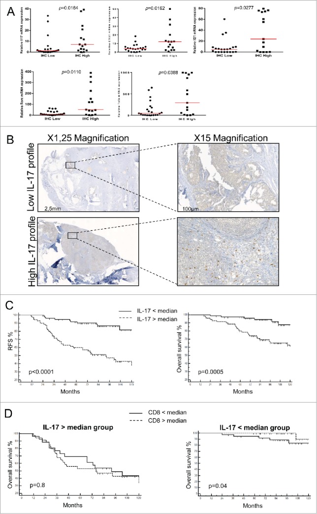
IL-17-infiltrating cells drive immunosuppression in human breast cancer/IL-17-infiltrating cells negatively affect human breast cancer prognosis. (A) IL-17 expression in 36 breast cancer tumor samples was determined by immunohistochemistry (IHC). Samples were then classified as IL-17high and IL-17low based on IHC results and the expression of IL17, ENTPD1, IL21, RORC and TNFA was determined using RT-qPCR. (B) Representative picture of IL-17+ staining of breast tumor using immunhistochemistry. We represent a tumor with a low (upper panel) and high (lower panel) IL-17 infiltrates. N = 145 patients. (C) Kaplan–Meier curves of relapse free survival (left panel) and overall survival (right panel) for patients with local breast cancer according to the presence of a high or low density of IL-17+ cells in the tumor stroma. N =145 patients. (D) Kaplan–Meier curves of overall survival according to the presence of a high or low density of CD8+ cells in the tumor stroma in the group of a high (left panel) or low (right panel) density of IL-17+ cells infiltrate. N = 70.