Figure 2.
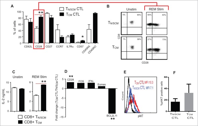
Effector cells derived from CD8+ TCM precursors expressed higher levels of CD28 and IL-2 production upon stimulation. Purified CD8+ TN/SCM and CD8+ TCM were subject to 14 d in vitro REM stimulation in the presence of rhIL-2 (50U/mL). (A) Phenotypic analysis of differentiated effector cells derived from CD8+ TN/SCM (TN/SCM CTL) and CD8+ TCM (TCM CTL) following expansion. Mean percentages of immunoreactive cells + SEM from 4 different donors are presented (**p < 0.01, *p < 0.05). (B) Representive flow cytometric analysis of CD28 on the CTLs derived from CD8+ TN/SCM and CD8+ TCM. (C) Supernatants were collected after overnight co-incubation of either unstimulated CD8+ TN/SCM and CD8+TCM or stimulated CD8+ TN/SCM and CD8+TCM with OKT3 expressing LCL. Cytokine levels (means +SEM of triplicate wells) were determined using cytometric bead array. **p < 0.01. Representative data of 4 experiments are depicted. (D) After 14 d of stimulation, RNA was extracted and analyzed for genes that confer differentiation, survival, and apoptotic qualities of effector T cells and 4 control genes including housekeeping genes, RT control and positive PCR control. Medians of fold change of CD28, FOS, IFNγ, Eomes and BCL2L11 mRNA from cells derived from CD8+ TCM (TCM CTL) vs. CD8+ TN/SCM (TN/SCM CTL) from 4 individual donors are presented. **p < 0.01. (E) Effector T cells derived from CD8+ TN/SCM (TN/SCM CTL) (blue) and CD8+TCM (TCM CTL) (red) were stained for intracellular phosphorylated AKT (pAKT). Fluorochrome conjugated isotype matched antibody stained cells are shown in black. (F) Percentages of AKT+ cells from 4 donors are presented.

