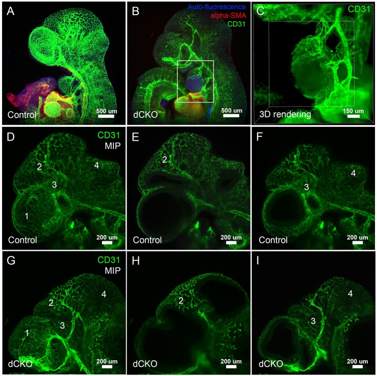
Fig. 4.
Severe vessel defects caused by deletion of Yap and Taz. Whole-mount CD31 immunofluorescence staining in a control (A) and a Wnt1Cre; Yap; Taz dCKO mutant (B) reveals vessel defects and endothelial-lined hemangiomas in the forebrain and mandible in Wnt1Cre; Yap; Taz dCKO mutants (B). (C) The boxed area in B at a higher magnification, focusing on the endothelial-lined hemangiomas in the branchial arch in the Yap; Taz dCKO embryo. Whereas control embryos have normal vessel development in brain (D-F), Wnt1Cre; Yap; Taz dCKO mutants have vessel regression and disorganization (different regions are labeled 1-4) (G-I). Endothelial cells are stained with CD31 antibody (green), smooth muscle cells are stained with SMA antibody (red) and auto fluorescence is blue.

