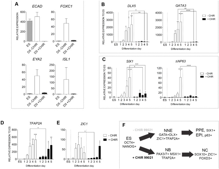
Fig. 3.
WNT restricts non-neural ectoderm fate in differentiating hESCs. (A) Quantitative gene expression analysis of placodal and/or surface ectoderm associated transcripts (ECAD, FOXC1, EYA2, ISL1) in ESCs (ES), D5+CHIR or D5−CHIR cultures. (B-E) Quantitative gene expression analysis in control ESCs, −CHIR and +CHIR cultures from day 1 to day 5, for competent non-neural ectoderm (DLX5 and GATA3; B), preplacodal ectoderm (SIX1; C), epidermal progenitor (ΔNP63; C) and prospective neural border (TFAP2A and ZIC1; D,E) genes. (F) Model depicting the stage-wise in vitro differentiation of placodal/epidermal ectoderm and NC cells. EPI, epidermal progenitors; NNE, non-neural ectoderm; PPE, preplacodal ectoderm. All panels normalized to ESCs except for ECAD in panel A, which is normalized to day 5 CHIR-treated cultures. Error bars represent s.e.m. Data were pooled from three or more independent experiments. *P≤0.05, **P≤0.01, ***P≤0.001, ****P≤0.0001.

