Fig. 2.
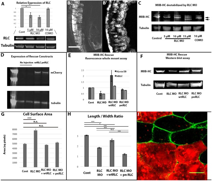
Regulatory light chain molecular and cellular phenotypes. (A) Western blotting reveals RLC protein decreases in a MO dose-dependent manner compared with COMO (n=13 gels). (B) Scattered COMO (B) or RLC morphant (B′) cells in intact stage 17 embryos visualized in 15 μm Z-projections of stacks by means of co-injected fluorescent Rhodamine-dextran reveals a cell shape dependence on RLC levels. (C) Myosin IIB heavy chain levels exhibit an RLC MO dose-dependent decrease associated with an increase in proteolytic degradation. (D) Injecting wtRLC-mCherry or pnRLC-mCherry mRNA into developing embryos leads to protein expression as detected by anti-mCherry antibody. (E,F) Two methodologies show that wtRLC and pnRLC expression rescues MHC-IIB stability; quantification of RLC and myosin IIB immunofluorescence levels in RLC morphant dorsal marginal zone explants (E; n=7 explants/condition) and western blot analysis of myosin IIB heavy chain levels in RLC MO embryos (F). (G) RLC morphant cells have a larger surface area than control cells in explants and this phenotype can be rescued by either wtRLC or pnRLC expression (n is at least 52 cells/condition). (H) However, RLC morphant cells have a reduced length-to-width ratio. Expression of wtRLC but not pnRLC substantially rescued this cell shape phenotype (n, at least 14 cells/condition). (I) Morphant cells expressing pnRLC labeled with Rhodamine-dextran (red) display lower length-to-width ratios than corresponding control cells (green). Error bars represent s.e.m. Cont, control. Scale bar: 100 μm in B and 20 μm in I.

