Fig. 3.
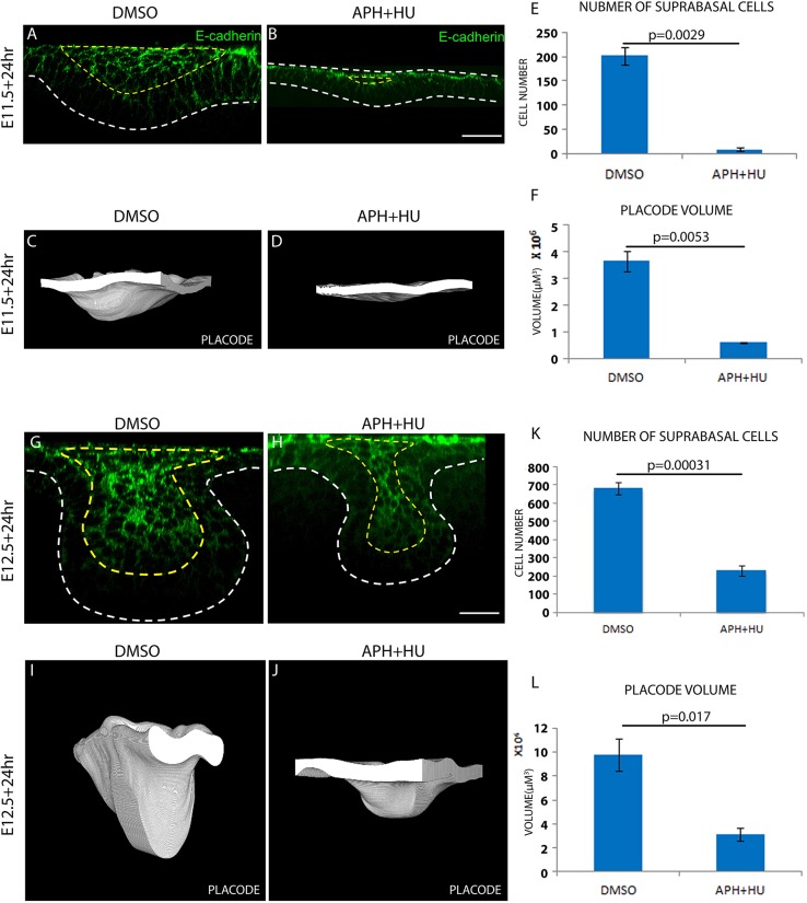
Cell proliferation controls suprabasal cell formation and the size of the early tooth. (A,B,G,H) E-cadherin (DECMA-1) immunofluorescence showing suprabasal cells of tooth placodes cultured from E11.5 (A,B) or E12.5 (G,H) with vehicle (DMSO) or proliferation inhibitors (APH+HU) for 24 h. (C,D,I,J) 3D rendering of tooth placodes cultured from E11.5 (C,D) or E12.5 (I,J) for 24 h with vehicle or proliferation inhibitors. White areas show the front-facing (virtually) cut face of the epithelium. (E) Quantification of suprabasal cell number in A and B. (F) Measurements of placode volume in C and D. (K) Quantification of suprabasal cell number in G and H. (L) Measurements of placode volume in I and J. For all quantifications, n=9 specimens (three from each of three litters). Error bars indicate s.d. Scale bars: 50 μm.

