Figure 4.
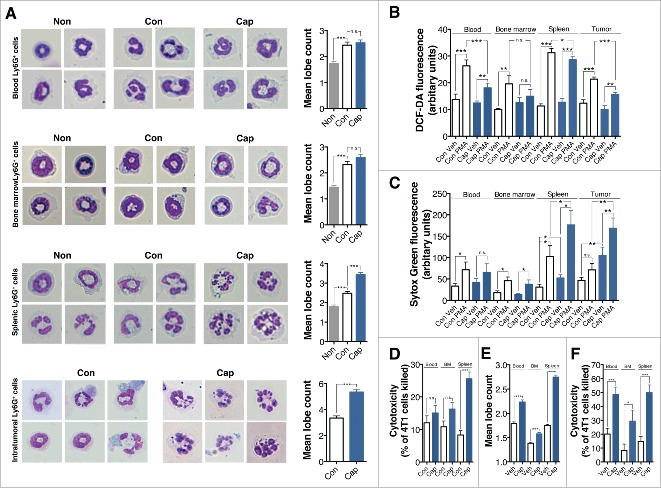
Captopril induces neutrophil hypersegmentation in a murine tumor model. (A) Left panel, representative images of Wright-Geimsa staining of Ly6G+ cells. Right panel, mean lobe counts of Ly6G+ cells. Non, Ly6G+ cells from naive mice; Con, Ly6G+ cells from control tumor-bearing mice; Cap, Ly6G+ cells from captopril-treated tumor-bearing mice. (B)–(C) Function of hypersegmented Ly6G+ cells. Ly6G+ cells from tumor-bearing control mice or captopril-treated tumor-bearing mice were isolated and exposed to PMA for 4 h. (B) PMA-induced ROS generation from Ly6G+ cells. (C) PMA-induced NETs formation. (D) Cytotoxicity of Ly6G+ cells against 4T1 cells. Ly6G+ cells from control tumor-bearing mice or captopril-treated tumor-bearing mice were isolated and exposed to 4T1 cells at a ratio of 10:1 for 18 h. (E) Effects of captopril on Ly6G+ cells from naive mice. Blood, bone marrow, and splenic Ly6G+ cells were isolated from naive mice and exposed to 500 μM captopril for 4 h, and then mean lobe counts were measured. Veh, vehicle-treatment. (F) Cytotoxicity of captopril-treated neutrophils against 4T1 cells. Blood, bone marrow, and splenic Ly6G+ cells were isolated from naive mice and exposed to 4T1 cells at a ratio of 10:1 for 18 h in the presence or absence of 500 μM captopril. n = 5–10 mice for each group. n.s. not significant; *p < 0.05; **p < 0.01; ***p < 0.001. All results are shown as means ± SEMs.

