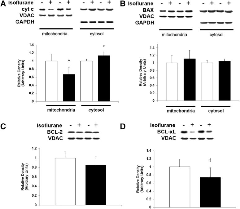
Figure 3.

Isoflurane induces cytochrome c release from mitochondria in the developing retina. Mitochondria and cytosol were isolated from retina immediately after 1-hour exposure to air (−) or isoflurane (+). Representative immunoblots for (A) cytochrome c (cyt c), (B) Bax, (C) BCL-2, and (D) BCL-xL are depicted. Voltage-dependent Anion Channel and glyceraldehyde 3-phosphate dehydrogenase were used as loading controls for mitochondrial and cytosolic protein, respectively. Graphical representations are shown. White bars are air-exposed controls and black bars are isoflurane-exposed cohorts. Air-exposed values were arbitrarily set to 1. Values are expressed as means + SD. N = 5–6. *P = 0.015 versus air-exposed cohort; 95% CI, 0.02–0.25. †P = 0.004 versus air-exposed cohort. ‡P = 0.033 versus air-exposed cohort; 95% CI, −0.54 to 0.02.
