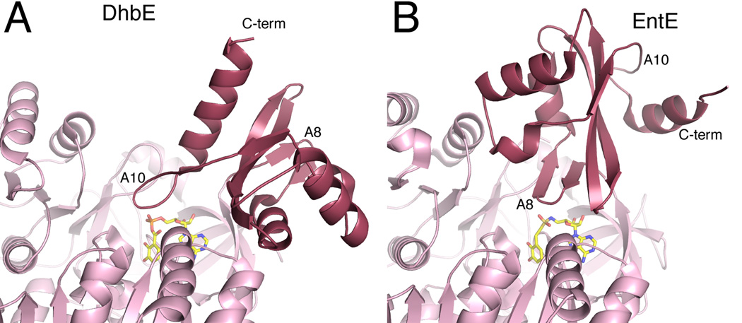
Figure 4.
Domain alternation of NRPS adenylation domains. The structures of two free-standing adenylation domains are shown from two bacterial siderophore synthesis, A. DhbE from the bacillibactin NRPS of B. subtilis and B. EntE from the enterobactin NRPS of E. coli. The DhbE structure (PDB 1MDB) is in the adenlyate-forming conformation, with the A10 motif of the C-terminal subdomain directed towards the active site. The EntE structure (PDB 4IZ6) adopts the thioester-forming conformation with the A8 motif near the active site. The carrier protein and the pantetheine cofactor of structure 4IZ6 are not shown for clarity.

