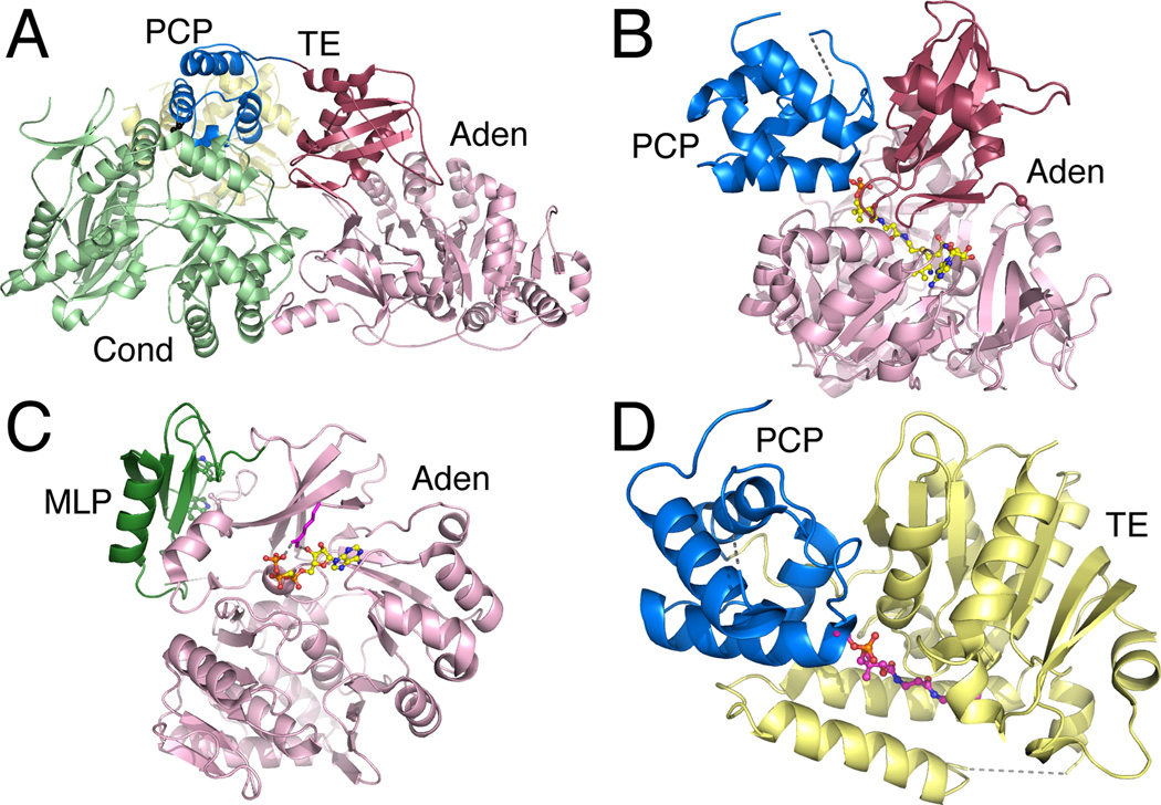
Figure 7.
Crystal Structures of multi-domain NRPS proteins. A. The structure of the complete termination module of SrfA-C (PDB 2VSQ) shows the PCP interactions with the downstream side of the condensation domain. B. The adenylation-PCP structure of PA1221 (PDB 4DG9) illustrates a functional interface between the PCP and the thioester-forming conformation of the adenylation domain. C. The structure of the SlgN1 protein (PDB 4GN5) shows the interaction between the MbtH-like domain (forest green) interacting with the N-terminal subdomain of the adenylation domain. The conserved tryptophan residues are highlighted in the MLP. D. The PCP-thioesterase domain of EntF (PDB 3TEJ) is shown illustrating the binding of the pantetheine into the active site of the thioesterase domain.

