Fig. 3.
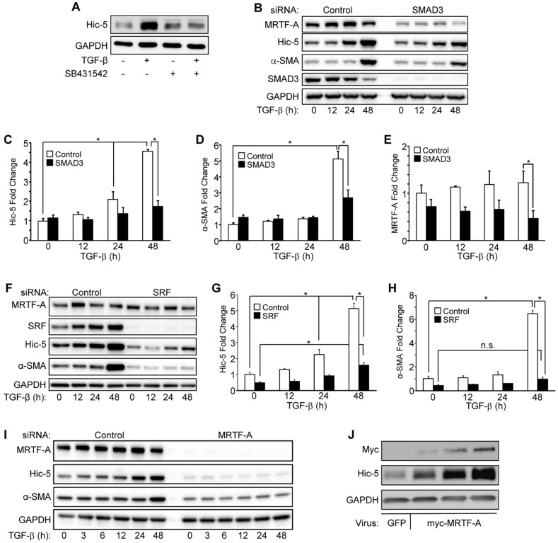
Induction of Hic-5 by TGF-β requires the SMAD3 and SRF–MRTF-A pathways. (A) NHDFs that had been treated with either 10 µM SB431542 (TGF-β receptor kinase inhibitor) or vehicle control with or without 10 ng/ml of TGF-β for 24 h were lysed and subjected to western blotting for Hic-5 and GAPDH (loading control) levels (representative blot, n=3 experiments). (B–E) NHDFs that had been treated with either non-targeting control or SMAD3-targeting (‘SMAD3’ on top of image) siRNAs were treated with TGF-β for the indicated times and analyzed by western blotting (B, representative blot) for Hic-5, α-SMA, SMAD3, MRTF-A and GAPDH. Data from three independent experiments were pooled, and the induction by TGF-β of Hic-5 (C) α-SMA (D) and MRTF-A (E) was quantified. (F–H) NHDFs that had been transfected with either SRF-targeting (‘SRF’ on top of image) or non-targeting siRNAs were stimulated with TGF-β for the indicated times and analyzed by western blotting (F, representative blot) for SRF, Hic-5, α-SMA, MRTF-A and GAPDH. (G,H, pooled data, n=3). (I) NHDFs that had been transduced with lentiviruses expressing MRTF-A-targeting (‘MRTF-A’ on top of blot) or non-targeting shRNAs and stimulated with TGF-β for 0, 3, 6, 12, 24 and 48 h (representative blot, n=3). NHDFs expressing MRTF-A-targeting shRNA exhibited reduced basal levels of Hic-5 (compare to t=0), and Hic-5 protein levels failed to respond to TGF-β stimulation at any time point. (J) NDHFs that had been infected with three different concentrations of adenovirus encoding MRTF-A or a high dose of adenoviral GFP. Statistical significance was determined by two-way ANOVA and Tukey's post-hoc analysis (*P<0.05). Error bars are +s.e.m. n.s., not significant.

