Fig. 2.
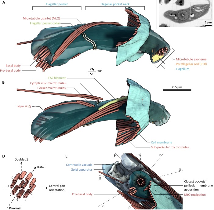
The organisation of the Leishmania mexicana flagellar pocket. (A) Lateral view of the flagellar pocket structure of a representative promastigote 1K1N L. mexicana cell (see C), as determined using serial section electron tomography. Flagellum, flagellar pocket and associated structures are shown, relative to a small portion of the pellicular microtubules and membrane. Generated using tomogram PL1. (B) The structure shown in A rotated 90° around the horizontal axis. (C) Low-magnification electron micrograph of the cell whose flagellar pocket is shown in A and B. Section 4 of the six used to build the tomogram is shown. The location of the nucleus (N), kinetoplast (K), flagellar pocket (FP) and region reconstructed using electron tomography (box) are indicated. (D) The standard axoneme doublet microtubule numbering scheme and corresponding coordinate system that we used to describe organelle locations relative to the flagellum within the flagellar pocket. (E) Anterior to posterior view of the base of the flagellar pocket shown in A and B. Five structures and/or organelles, which were found in a consistent position relative to the flagellum and flagellar pocket in all tomograms analysed, are indicated, mapped relative to the axoneme doublet numbering scheme. Generated using tomogram PL1.

