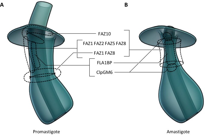
Fig. 7.
Possible FAZ protein locations in the pocket ultrastructure. 3D cartoon of possible protein localisations, inferred from light micrographs of eYFP fusions of FAZ proteins and the pocket structure derived from electron tomography analysis. (A) The promastigote flagellar pocket. (B) The amastigote flagellar pocket.

