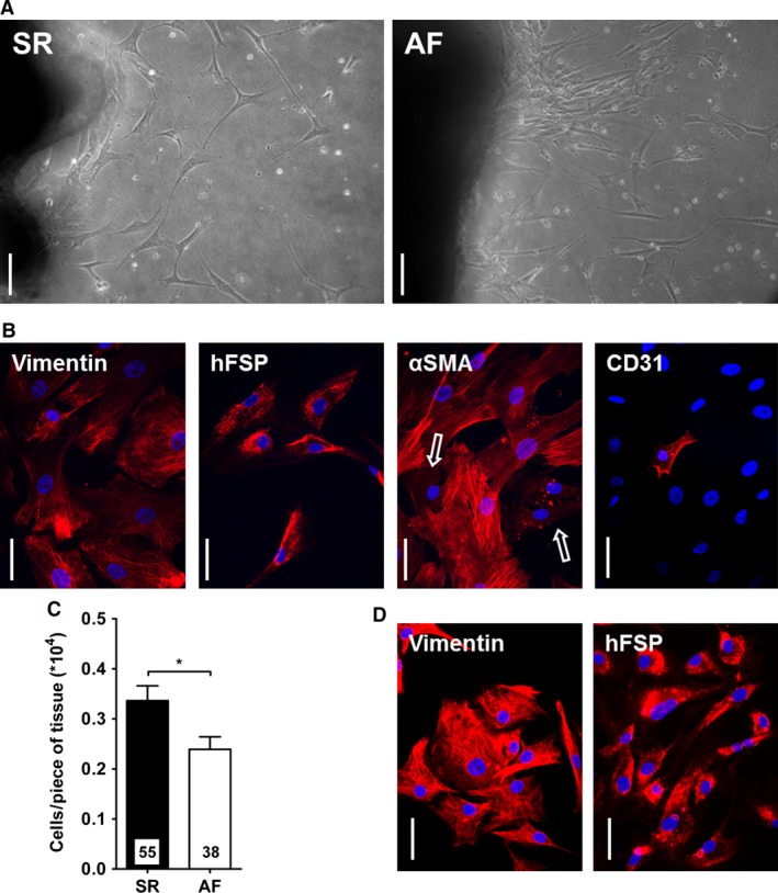
Figure 1.

Characterization of cultured fibroblasts. (A) Fibroblasts growing out of explants from SR and AF samples after 1 week of culture. Scale bar: 100 μm. (B) Staining for the expression of vimentin, human fibroblast surface protein (hFSP), alpha‐smooth muscle actin (α SMA) and CD31 in AF fibroblasts before replating. Nuclei are stained in blue with DAPI. Cells expressing α SMA‐positive myofilaments are easily distinguished from negative cells (arrows). Scale bar: 50 μm. (C) Cell count after 3 weeks of culture. The number of cells was normalized to the number of tissue pieces. *P < 0.05, unpaired Student's t‐test. (D) Staining for Vimentin and hFSP in AF fibroblasts after replating. Nuclei are stained in blue with DAPI. Scale bar: 50 μm.
