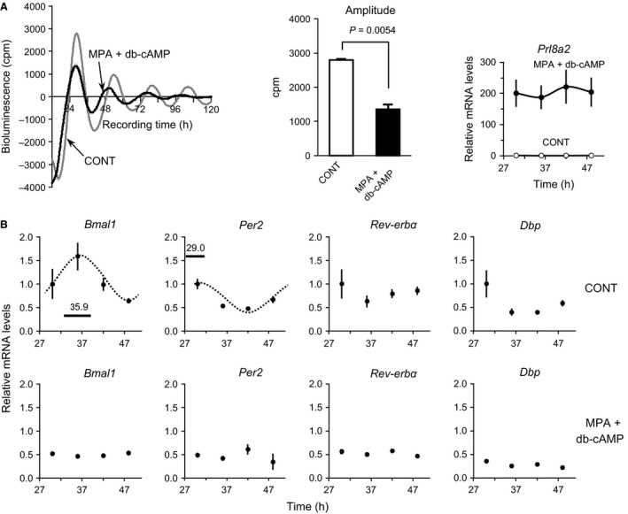
Figure 3.

Per2 oscillation profiles and expression levels of core clock genes in UESCs during in vitro decidualization. (A) UESCs were isolated from the uterine horns on day 4.5 of pregnancy and cultured for 3 days prior to any treatment. Decidual cells were obtained by treating confluent UESCs for 4.5 days with 0.1 mmol/L MPA plus 0.5 mmol/L db‐cAMP. Both types of cells were synchronized with DXM for 2 h prior to monitoring. The amplitude of Per2‐dLuc oscillations was determined by the single Cosinor method. The mRNA level of Prl8a2, a marker for UESCs decidualization, was analyzed in the control and decidual cells by RT‐qPCR. (B) According to the first Per2‐dLuc phase in panel A, total RNA samples were collected from control and decidual cells at the indicated times after synchronization and RT‐qPCR analyses of transcript levels were performed. The transcript levels were calculated and normalized to each value given by the control sample at 30 h. The Cosinor analysis method was used to determine the rhythmic expression of examined genes. Statistical rhythmicity (dotted curves, P < 0.05) and peak time with 95% confidential intervals (line length) are shown in each panel.
