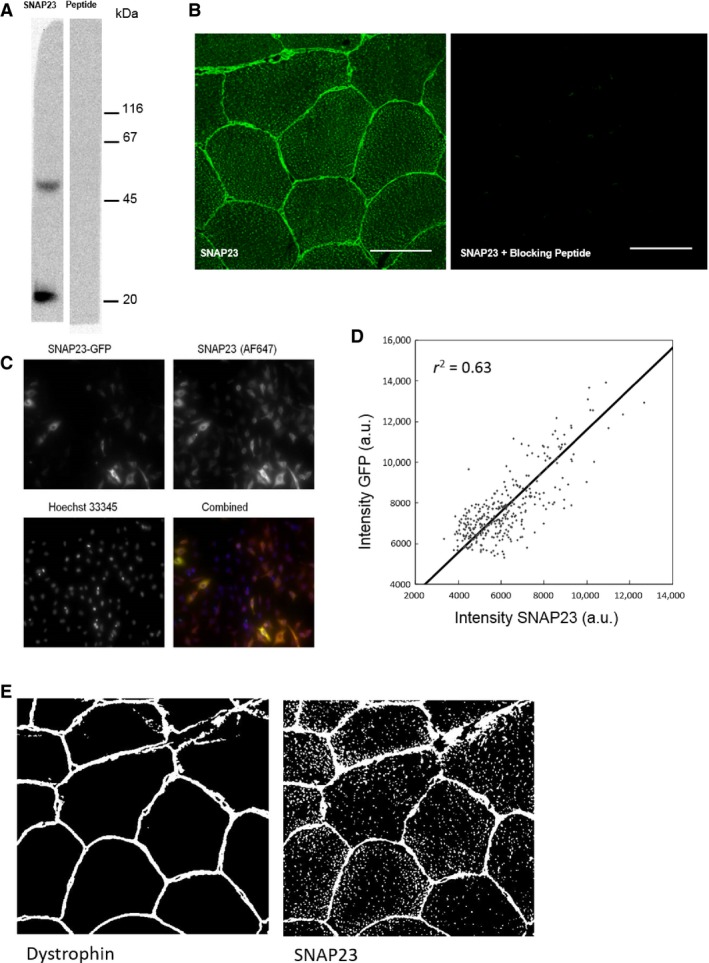
Figure 1.

Antibody validation. (A) Immunoblot using anti‐SNAP23 (Synaptic Systems) of a homogenate of human skeletal muscle from a lean male. The molecular weight of SNAP23 is 23 kDa. The band with the higher molecular weight is likely to be a SNARE complex containing at least one molecule of SNAP23. Coincubation of anti‐SNAP23 with blocking peptide eliminated both bands from the blot. (B) Immunostaining of SNAP23 is shown as well as immunostaining using anti‐SNAP23 preincubated with an excess of blocking peptide. Almost all signals are eliminated. Bar = 50 μm. (C) HeLa cells transfected with 100 μg SNAP23‐GFP plasmid incubated for 48 h. The cells displayed a heterogenous expression of the plasmid and show colocalization of SNAP23‐GFP and immunostaining for SNAP23 with anti‐SNAP23 (1:100 dilution). In the merged image the SNAP23‐GFP plasmid is visualized in green, SNAP23 stained with anti‐SNAP23 in red, nuclei in blue, and colocalization of SNAP23‐GFP with anti‐SNAP23 in yellow. Bar = 100 μm. (D) Quantification of the cytoplasmic fluorescence intensities of SNAP23‐GFP and immunostaining with anti‐SNAP23. Data points are obtained from 337 cells in three replicate wells and show a linear relationship (r 2 = 0.63). (e) Example binary “masks” for the plasma membrane and SNAP23 showing the masks used during the colocalization analysis to identify the “objects” in the field of view (i.e., SNAP23, mitochondria, lipid droplets, or plasma membrane). Bar = 50 μm.
