Figure 3.

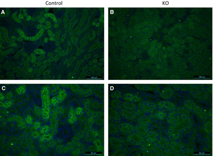
Immunostaining of kidney sections with anti‐AGT antibody (green) from control (A and C) and PRR KO mice (B and D). Images are representative of three different mice. (A and B) are 200× and (C and D) are 400× magnification.

Immunostaining of kidney sections with anti‐AGT antibody (green) from control (A and C) and PRR KO mice (B and D). Images are representative of three different mice. (A and B) are 200× and (C and D) are 400× magnification.