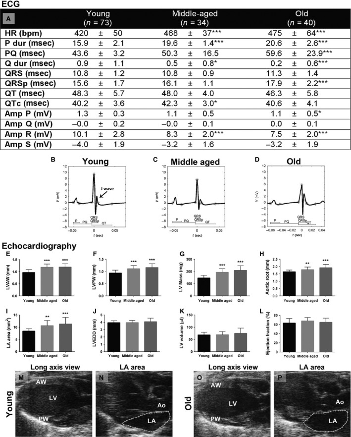
Figure 1.

ECG and echocardiographic results of young (2–3 months old, n = 73), middle‐aged (12–14 months old, n = 34), and old (20–24 months old, n = 40) C57Bl/6J mice. ECG measurements of the lead II (A) and representative ECG of young (B), middle‐aged (C), and old (D) mice with appropriate ECG intervals marked. Echocardiographic measurements of LVAW thickness (E), LVPW thickness (F), LV mass (G), aortic root diameter (H), LA area (I), LVEDD (J), LV volume (K), and ejection fraction (L). Representative long‐axis view B‐Mode images of young (M, N) and old (O, P) mice. HR, heart rate; P dur, duration of P wave; Q dur, duration of Q wave; Amp, amplitude; LV, left ventricle; LVAW, LV anterior wall; LVPW, LV posterior wall; LVEDD, LV end‐diastolic diameter; LA, left atrium. Results are expressed as mean ± SD. One‐way ANOVA with Dunnett's post hoc test was used, *P < 0.05, **P < 0.01, and ***P < 0.001 compared with the group of young mice.
