Fig. 1.

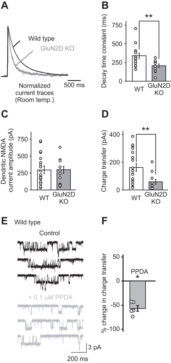
N-methyl-d-aspartate (NMDA) receptors that contain GluN2D subunits are expressed in the soma and dendrites of stellate cells. A: dendritic NMDA receptor currents were evoked by a single-burst stimulation of parallel fibers (PFs) at 100 Hz (4 stimuli) and recorded at +40 mV at room temperature. NMDA receptor currents recorded in stellate cells from GluN2D knockout (KO) mice exhibited a faster decay time compared with wild-type (WT) mice, as shown in the normalized currents (GluN2 KO, gray trace; WT, black trace). B: summary of the decay time constants of NMDA receptor currents recorded in stellate cells from WT and GluN2D KO mice. Deletion of GluN2D accelerated the decay time of evoked dendritic NMDA receptor currents. C: the amplitude of dendritic NMDA receptor-mediated currents. D: the charge transfer of dendritic NMDA receptor-mediated currents, which was calculated as the time integral of NMDA receptor currents, was reduced in GluN2D KO mice. Thus NMDA receptors that contain GluN2D subunits are present in cerebellar stellate cells. Open circles represent data from individual cells. E: outside-out somatic patches were excised from stellate cells from WT mice, and NMDA receptor currents were recorded during the application of 10 μM NMDA and 10 μM glycine. PPDA [(2S*,3R*)-1-(phenanthren-2-carbonyl)piperazine-2,3-dicarboxylic acid; 0.1 μM] was added to the superfusion medium to block GluN2D-containing NMDA receptors. F: PPDA reduced the charge transfer of somatic NMDAR currents. *P < 0.05. **P < 0.01.
