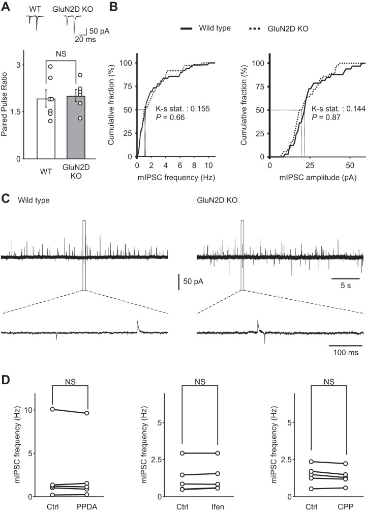
Fig. 4.
NMDA receptors do not regulate basal spontaneous GABA release in cerebellar stellate cells. A, top: representative recordings of excitatory postsynaptic currents (EPSCs) evoked by PF stimulation with two consecutive stimuli in WT (left) and GluN2D KO mice (right). Bottom: the paired-pulse ratio of EPSCs at the PF-stellate cell synapse (the ratio of amplitude of EPSC2/EPSC1) was not modified in GluN2D KO mice (WT n = 7, GluN2D KO n = 6), indicating that the release probability of glutamate from PFs was not altered. B: cumulative distribution of mIPSC frequency (left) and amplitude (right) in WT (solid line) and GluN2D KO mice (dashed line). mIPSC frequency was not altered in GluN2D KO mice (n = 36) compared with WT mice (n = 49). Thus deletion of GluN2D did not change basal spontaneous GABA release from stellate cells. C: representative traces of mIPSCs (outward currents) in WT (left) and GluN2D KO (right) mice. The bottom traces are enlargements of regions designated by the dashed lines. D: NMDA receptor blockers did not alter basal mIPSC frequency recorded at −60 mV using a CsCl-based pipette solution in WT stellate cells. After a stable baseline was obtained in the presence of 0.5 μM TTX and 5 μM 2,3-dihydroxy-6-nitro-7-sulfamoyl-benzo[f]quinoxaline-2,3-dione to isolate mIPSCs, PPDA (left, 0.1 μM, n = 5), ifenprodil (center, 3 μM, n = 5) or 3-[(R)-2-carboxypiperazin-4-yl]-propyl-1-phosphanoic acid (R-CPP; right, 0.2 μM, n = 5) were added to the artificial cerebrospinal fluid for at least 20 min.

