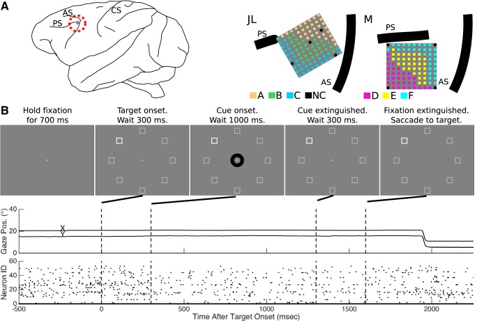
Fig. 1.
Experimental setup. A: microelectrode arrays were implanted in the left hemisphere, anterior to the knee of the arcuate sulcus (AS) and caudal to the posterior end of the principal sulcus (PS). The central sulcus (CS) is also indicated. Each recording session collected data from a block of 32 electrodes. For each monkey, electrode sites are colored according to membership in a block (A, B, or C for monkey JL; and D, E, or F for monkey M; NC, not connected). B: in each trial, the monkey maintained fixation through the presentation of the target stimulus until the central fixation point extinguished at which time the monkey made a saccade to the target. Example behavioral eye position data and neural spiking data for a single trial are presented in the middle and bottom panes, respectively. Note that the screen y-axis origin was at the top of the screen, and down was positive.

