
Fig. 2.
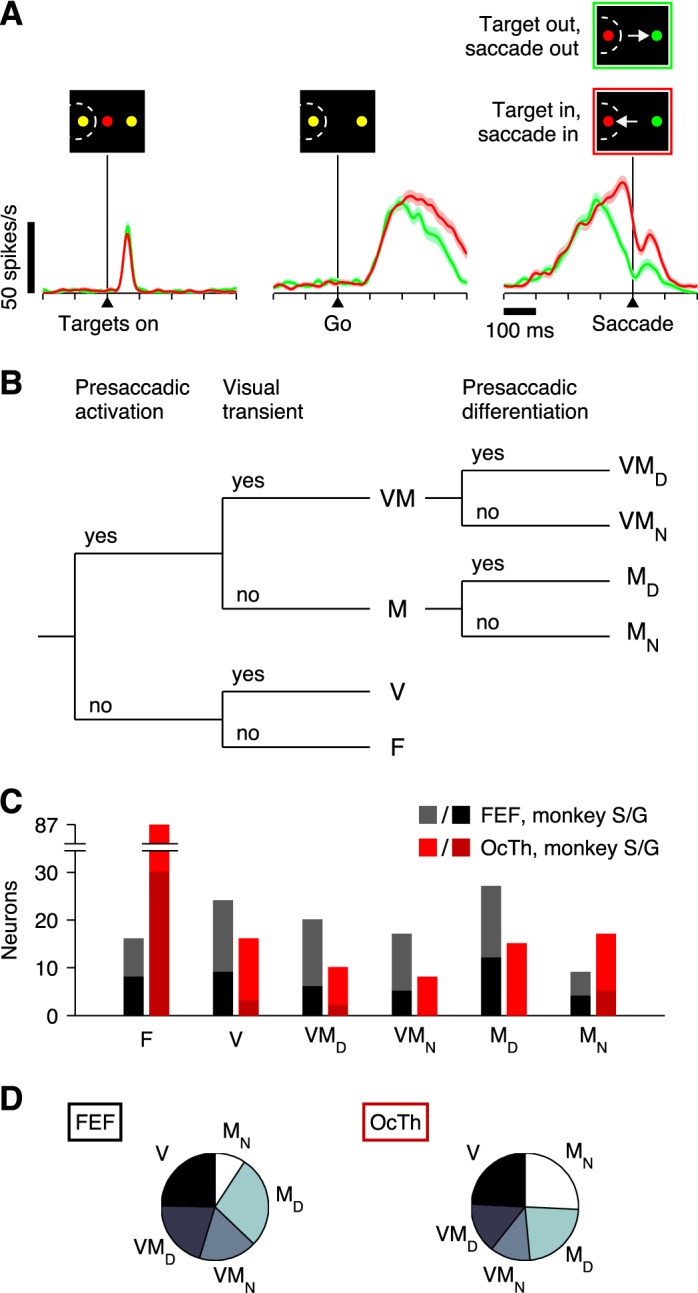
Neuronal classification. Six neuronal categories were defined based on the activity evoked during the CS task. A: example of a single OcTh oculomotor thalamus (OcTh) neuron recorded in the CS task. Traces show mean firing rate as a function of time for correct responses into (red) or away from (green) the response field (RF), with spike trains aligned on the onset of the choice targets (left), on the go signal (middle), and on saccade onset (right). Shaded areas indicate ± 1 SE across trials. All processing times were included. Note visual response following targets on, increase in firing before the saccade (i.e., activation), and differentiation between choices into and away from the RF. This unit was classified as visuomotor differential. B: decision tree indicating the 3 classification criteria used (top) and resulting neuronal categories: F, flat; V, visual; VMD, visuomotor differential; VMN, visuomotor nondifferential; MD, motor differential; and MN, motor nondifferential. In addition, the VM and M labels refer to visuomotor and motor groups, respectively, regardless of their differentiation. C: numbers of neurons of each type recorded in frontal eye field (FEF; black/gray bars) and OcTh (red bars). Light and dark shades indicate data from monkeys S and G, respectively. D: proportions of responsive neurons in FEF (left) and OcTh (right). Plots are based on the data in C with the F category excluded.