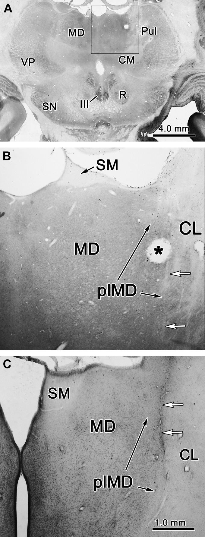
Fig. 3.
Localization of thalamic recording sites. A: representative, low-magnification section from monkey S, stained for cytochrome oxidase activity. B: high-magnification view of the region inside the square in A. *Electrolytic microlesion made after a recording session. White arrows indicate electrode tracks. C: Nissl-stained section from monkey G. White arrows indicate gliosis around an electrode track. Same scale as in B. CL, central lateral nucleus; CM, centromedian nucleus; III, oculomotor nucleus; MD, mediodorsal nucleus; plMD, paralamellar mediodorsal nucleus; Pul, pulvinar; R, red nucleus; SM, stria medullaris; SN, substantia nigra; VP, ventral posterior nucleus.

