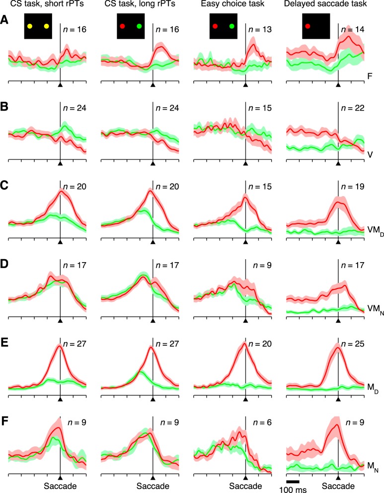
Fig. 6.
Presaccadic activity in FEF across tasks. All traces correspond to instantaneous firing rate as a function of time for correct responses into (red) or away from (green) the RF. Each trace is an average across all neurons recorded in each condition (n, indicated in each plot), where each neuron's contribution was divided by its overall maximum response (normalization factor; see materials and methods). Thus the y-axes represent normalized activity going from 0 (no firing) to 1 (maximum firing). Shaded areas indicate ±1 SE across neurons. All data are aligned on saccade onset. Columns correspond to different tasks, as marked at the top. A–F: responses for 6 neuron types, F (A), V (B), VMD (C), VMN (D), MD (E), and MN (F), as indicated on the right.

