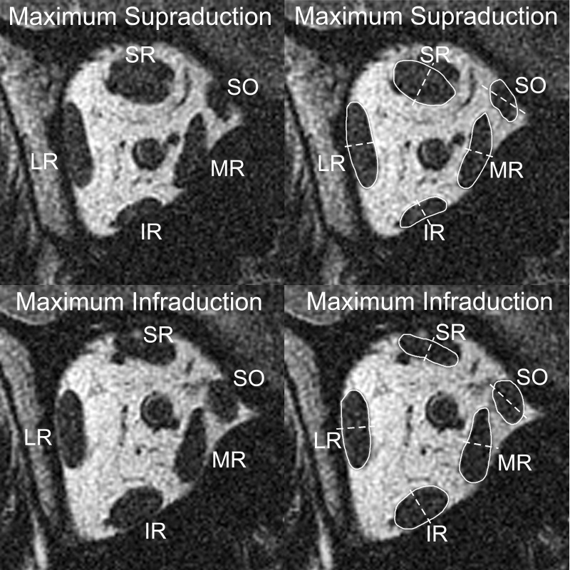
Fig. 3.
MRI at plane −5 (10 mm posterior to the globe-optic nerve junction) in maximum supraduction (top) and maximum infraduction (bottom). Images on right are duplicates with the extraocular muscle bellies outlined in white and compartmental bisections marked with dashed white lines. From maximum supraduction to maximum infraduction, the medial half of the IR belly exhibited a larger increase in cross section than the lateral half. Likewise, the superior and inferior halves of the MR belly and the medial and lateral halves of the tilted SO belly demonstrate asymmetric morphological changes.

