
Fig. 2.
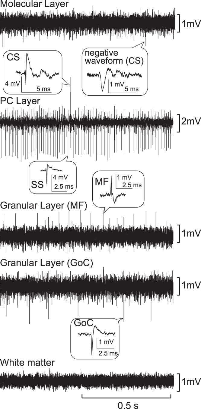
Identification of the different layers of the cerebellar cortex based on characteristic patterns of extracellular field potentials and single-cell activities. Typical field potential recordings for each layer of the cerebellar cortex are shown in order from surface to depth. Typical examples of single-unit activities are shown with higher time resolutions in the balloons. CS, complex spike; SS, simple spike.