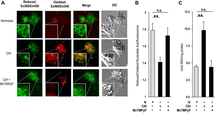
Fig. 4.
Effect of CIH on reactive oxygen species (ROS) levels in rat glomus cells. A: autofluorescence of reduced [excitation (Ex) λ366 nm/emission (Em) λ450 nm] and oxidized nucleotides (excitation λ460 nm/emission λ540 nm) was monitored as an index of ROS in rat glomus cells. Fluorescent and differential contrasting (DIC) photomicrographs of glomus cells treated with normoxia (top), CIH (middle), or CIH + manganese (III) tetrakis (1-methyl-4-pyridyl) porphyrin pentachloride (MnTMPyP; bottom). Squares in each photomicrograph represent magnified (2×) images of the region denoted by dotted lines. B: quantitative analysis of the ratios of reduced/oxidized nucleotide autofluorescence intensity (means ± SE) in glomus cells treated with normoxia (n = 42 cells), CIH (n = 58 cells), or CIH and MnTMPyP (n = 49 cells). C: malondialdehyde (MDA) levels, representing the oxidized lipids, were analyzed as an index of ROS in carotid bodies from rats treated with normoxia, CIH, or CIH and MnTMPyP. Rats were treated every day for 10 days with MnTMPyP (5 mg/kg ip) before a daily regimen of CIH treatment. Data represent means ± SE from 3 independent experiments (4 carotid bodies/experiment). **P < 0.01; n.s., not significant (P > 0.05).

