Fig. 9.
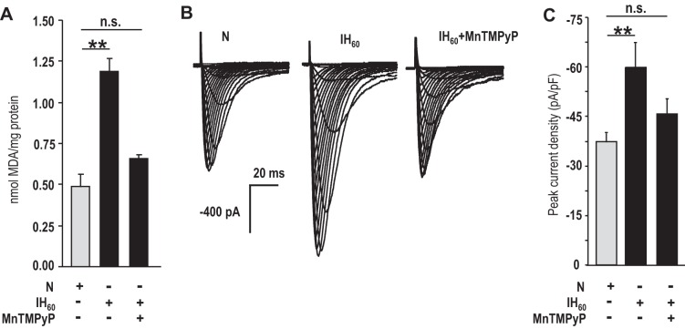
ROS mediates the augmented Ca2+ current in IH-treated CaV3.2-HEK cells. A: MDA levels were determined as an index of ROS generation. Average data (means ± SE) of MDA in CaV3.2-HEK cells treated with normoxia or IH60 or IH60 in the presence of 50 μM MnTMPyP (n = 3 independent experiments each). B: representative Ca2+ currents from CaV3.2-HEK cells treated with normoxia or IH60, with and without 50 μM MnTMPyP. The recording protocols were same as in Fig. 8. C: average (means ± SE) normalized peak current density obtained with a step to −30 mV for normoxia (n = 9 cells)-, IH60 (n = 7 cells)-, and IH60 + 50 μM MnTMPyP (n = 9 cells)-treated CaV3.2-HEK cells. **P < 0.01; n.s., not significant (P > 0.05).

